Have you ever had braces? My dentist once told me that I had a big mouth (it was insulting at first, but she told me it was a good thing), so my teeth had plenty of room to move into place. If you’re one of the many, many unlucky ones to have had braces, you can thank the system of tissues that anchor your teeth to the jaws for giving them the ability to move over time. Your teeth – unlike those of many reptiles and fish – are able to move over time, because they are not fused to your jaws. Instead of acting like a tent peg, drilled into the ground and fixed in place, your teeth are suspended by a network of ligaments, like a tarp hanging over your tent (to continue the camping metaphor). This means that teeth maintain a flexible, somewhat moveable connection to the jaws that can cope with the changes and forces that occur to a mammal’s teeth over time. Braces take advantage of this by pulling teeth into better positioning and making the ligaments stretch and re-attach themselves as the teeth move.
The tissues that make the connection between the teeth and the jaws collectively form the periodontium (Perio- meaning “around”, and dontium– refers to the teeth). In mammals, the main part of the periodontium is the periodontal ligament, which acts like a system of tiny bridge cables to suspend the tooth within its socket. The anchoring points for the ligament along the root are in the cementum, a bone-like tissue that coats our tooth roots. On the other end is the alveolar bone, the bone layer that forms our tooth sockets and anchors the ligament to the jaws. Because the ligament is a soft tissue, it doesn’t show up in your X-rays from the dentist’s office and it isn’t preserved in fossil mammal teeth. Instead, teeth seem to be floating mysteriously in the tooth sockets in X-rays and fossils, but trust me, the ligaments should be there!
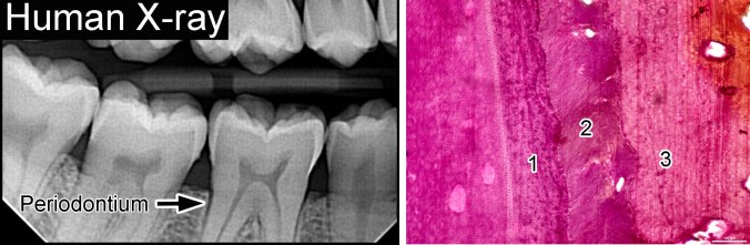
Human X-ray photo showing the periodontium (left). (Right) a closeup of a mammal periodontium on a microscope slide, showing the cementum coating the tooth root (1), the periodontal ligament (2), and the alveolar bone (3).
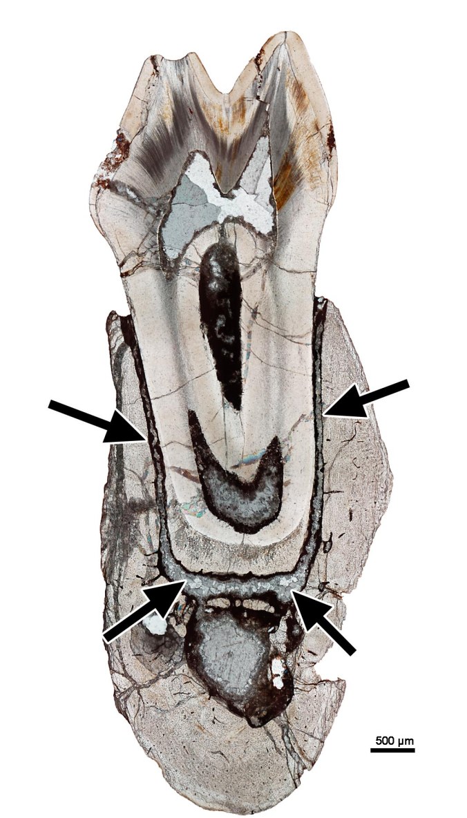
A thin-section through a jaw of a fossil mammal. The periodontal ligament is long gone, but the tooth floats in its socket, because the space where the ligament used to be has filled in with mineral (black arrows).
The periodontium is a remarkably complex system, but it has a number of benefits for mammal teeth. First, it gives teeth a way to shift around in the jaws as new teeth are added to the dentition. This is important for mammals, because the teeth need to maintain precise positioning so that they can come together to chew food, so being able to shuffle over and make room is essential. The second advantage is that the tooth is supported by a springy connection, giving each tooth a bit of give as opposing teeth come together. This cushioning effect helps the tooth last a lot longer.
Curiously, this complex tooth attachment is not unique to mammals. Despite seeming to be the “perfect” system to hold mammal teeth in place, it shows up in some odd groups. One of these groups is the Crocodilia (crocodiles, alligators, and their kin). Why it shows up in them is still a mystery, but the periodontia (the plural of “periodontium”) in mammals and crocodilians are virtually identical, and so they are treated together here.
What does the periodontium look like?
The most notable feature is the periodontal ligament. This ligament is made of tiny cables of protein called collagen. The ligament contains a number of cells, blood vessels, and nerves that help maintain it for the entire life of the tooth. The nerves within the ligament act like tiny pressure sensors, letting you know when you’re biting down hard on food (remember, there are no nerves along the tops of your teeth in the enamel, so it is the ligament that gives that feeling to your teeth).
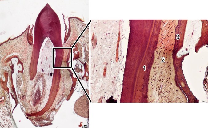
The periodontium in a caiman (a crocodilian). The tooth is suspended by the periodontal ligament when viewed in thin-section (left). (Right) a closeup of the three parts of the periodontium: the cementum (1), the periodontal ligament (2), and the alveolar bone (3). Note the tiny dark dots in the ligament, cementum and bone. Those are the nuclei of the cells!
These ligament fibers have to anchor into the roots of our teeth in order to have any chance of holding them in place when we eat. For this reason, we have cementum. Cementum comes in two types: acellular, and cellular. As the names suggest, cementum can have cells embedded in it, or not. In humans, the acellular cementum is the primary anchoring point for the ligament. This isn’t always the case in other animals. In other mammals and in crocodilians, the cellular cementum is the anchoring point for the ligament.

Cementum in a human tooth. The acellular cementum is the main site of attachment for the periodontal ligament (top right). Under cross-polarized light, the anchoring points for the ligament are visible (black arrows). (Bottom right) the other kind of cementum contains cells (white arrows) and in humans, forms a minor part of the periodontium.
One of my favourite things about cementum is that you can TELL what it does just by looking at it under the microscope. Though not always visible under normal light, with cross-polarizing filters, the anchoring points for the ligament in the cementum are visible.

A closeup of the root of a caribou tooth. Under normal light, the periodontal ligmament (2) and cementum (1) are visible, but under cross-polarized light, the cementum REALLY shines. That’s because this light filter highlights the anchoring fibers of the ligament that are embedded into the cementum (black arrows).
The other end of the periodontium is the tooth socket itself, which is made of a special bone layer called alveolar bone. Alveolar bone is NOT jawbone, but a bone layer that is actually made by your teeth (see below about a cool experiment that figured this out). This bone layer is the anchoring point for the periodontal ligament into the jaws. Alveolar bone is also found in between your teeth, giving the ligament something to anchor into. This is an incredibly dynamic tissue that can be eroded away by bone-eating cells (osteoclasts) and then re-made by bone-producing cells (osteoblasts) in order to progressively move a tooth through the jaws. I also learned the hard way that if you don’t floss regularly, the alveolar bone could start to recede, giving your ligament less of a foothold and creating room for infection. So…floss regularly!

A section through the jaw of a fossil mammal, showing the extent of the alveolar bone in between teeth.
How does the periodontium form?
When most people think of teeth, they think of enamel and dentine, the two hard tissues that make the body of the tooth. Cementum, the periodontal ligament, and the alveolar bone are rarely mentioned in studies of teeth, but they are incredibly important to our understanding of dental evolution and development. Some very cool experiments have shown us that the periodontium is actually part of the tooth, not the jaws. So how did they figure that out?
In 1971 Faculty of Dentistry researchers from the University of Toronto, Ten Cate, Mills, and Solomon, published a study on the development of the periodontium in lab mice. They wanted to know how a tooth forms its attachment to the jaws and whether the jawbones are involved in this process, or if the tooth does it by itself. In order to figure this out, they devised an experiment where they removed developing teeth in baby mice and transplanted them to different parts of the body (mostly along the back of the mouse, just under the skin).
This may sound like something out of science fiction, but in order to eliminate the role of the jaws in their experiment, this was the best option. And what did they find? After only a few weeks, the teeth developed enamel, dentine, AND the three parts of the periodontium! In other words, the tooth MADE its own tooth socket, out of bone, anchored into it with a periodontal ligament, and developed root cementum to complete the attachment…on the animal’s back! Some of the teeth had even started to move towards the surface, getting ready to erupt. This experiment raises some pretty horrific mental images, but it was profound, because it showed that teeth build their OWN periodontium, whether the teeth are in the jaws or not.
These types of transplant experiments have been repeated again and again, with the result always being the same: teeth develop enamel, dentine, and the periodontium regardless of the host tissue. The important thing to take away from this is that a tooth and its periodontium are a package deal. The cells forming each tooth make dentine, enamel, AND the periodontium. Each tooth is a highly integrated unit that forms a pre-set number of tissues every time, regardless of where it starts to form. Pretty cool!
What does the periodontium look like in other animals?
The periodontium in mammals is very complex, but we’re starting to understand how it forms and functions. By comparison, our knowledge of the periodontium in other vertebrates is quite poor. This has been an important facet of my research: trying to understand (a) how mammals evolved their periodontium; and (b) why it also shows up in several other groups, including crocodilians. I won’t devote any more time to that question just yet, but I’ll return to this subject in a later post with pictures of the periodontium in everyone’s favourite group: the dinosaurs!
If you enjoyed this blog entry and have a tooth-related question for me to tackle in a future entry, post a comment, send me an email at arl@ualberta.ca, or tweet me (@AaronLeBlanc6)
Some of the information presented here I got from these great resources on teeth:
Nanci, A. (2007). Ten Cate’s Oral Histology: Development, Structure, and Function, 7th ed. Mosby, St. Louis.
Osborn (1981). Dental Anatomy and Embryology: A companion to dental studies. Vol. 1. Book 2. Blackwell Scientific Publications.
Ten Cate, A.R., Mills, C., and Solomon, G. (1971). The development of the periodontium. A transplantation and autoradiographic study. Anatomical Record 170:356–380.
Ten Cate, A.R., Mills, C. (1972). The development of the periodontium: the origin of alveolar bone. Anatomical Record 173:69–77.
Ten Cate A.R. (1997). The development of the periodontium—a largely ectomesenchymally derived unit. Periodontology 2000 13: 9–19.
All of the images are my own.
